Patellar luxation is a common orthopedic condition in dogs, characterized by the displacement of the kneecap (patella) from its normal position in the trochlear groove of the femur, resulting in instability of the knee joint. This condition may be congenital or traumatic, the congenital form being most often seen in small breeds. Studies indicate that medial patellar luxation is more prevalent in females and causes lameness of varying intensity. Breeds such as the Poodle, Yorkshire Terrier, Shih Tzu, Dachshund, Pekingese, Lhasa Apso, Pomeranian, Chihuahua, Bichon Frise and Pug are particularly predisposed, while in large dogs the condition is more common in the Cocker Spaniel, Labrador Retriever, English Bulldog and Golden Retriever.
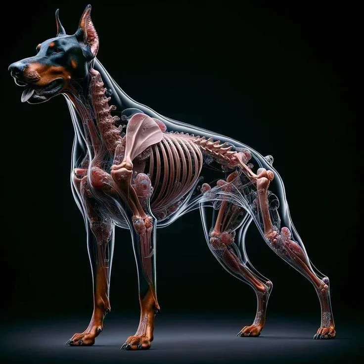
Patellar luxation occurs because of abnormalities in the development of the limb that misalign the quadriceps mechanism, made up of the quadriceps muscle, the patella and the patellar ligament. The associated anatomical abnormalities include lateral torsion of the distal femur, medial displacement of the quadriceps muscle group, lateral bowing of the distal third of the femur, dysplasia of the femoral epiphysis, rotational instability of the knee joint and tibial deformity. Congenital luxation is believed to have a strong hereditary component, probably polygenic and multifactorial. For this reason, it is recommended that animals be examined and graded through patellar luxation reports, so that matings can be planned to reduce the problem in breeds where it is very common. This way, the breeder can be legally assured of having taken the proper steps to avoid the problem, without acting in bad faith or recklessly in breeding the dogs.
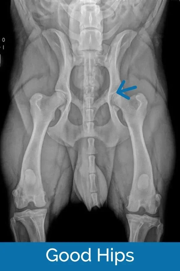
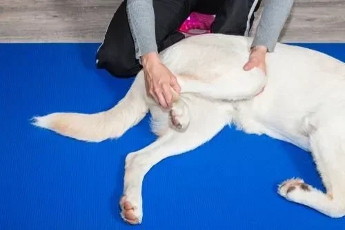
It is not possible to guarantee health regarding patellar luxation, just as with hip dysplasia, because it is a polygenic and multifactorial condition, but it is possible to prove that healthy dogs were used for breeding. Clinical signs vary with the severity of the luxation, ranging from no clinical signs, even in more severe grades, to intermittent or continuous lameness, pain, reluctance to walk or to climb onto beds, sofas and stairs, and abnormal swelling of the affected limb. Some dogs also show an atypical posture when walking, such as stretching the leg backward. In more severe cases, the difficulty moving can be significant. Diagnosis is based on the history, physical exam and imaging. During the clinical exam, the veterinarian assesses the stability of the patella and identifies signs of pain or discomfort. X-rays help confirm the diagnosis in more severe cases and detect associated bone deformities, such as periarticular osteophytes indicating osteoarthritis. Treatment may be conservative or surgical, depending on the grade of the luxation and the intensity of the signs. Conservative management is indicated for grade 1 or 2 luxations, especially in dogs with no or mild symptoms, and includes weight control, physical therapy to strengthen the muscles and changes in activity to avoid overloading the joint. Surgical treatment is recommended for grade 3 or 4 luxations or for cases in which conservative management does not relieve the signs.
Surgical techniques aim to realign the quadriceps mechanism and stabilize the patella in the trochlear groove. Among the most commonly used procedures are tibial tuberosity transposition, deepening of the trochlear groove and reconstruction of the soft tissues around the patella. The post-surgical prognosis is generally favorable for grade 1 to 3 luxations, with most dogs returning to normal use of the limb. However, in grade 4 luxations the prognosis may be more guarded, requiring a longer rehabilitation period. Patellar luxation is a condition that can compromise dogs' quality of life, so it is essential for breeders and owners to recognize the clinical signs early and seek proper treatment. Early diagnosis and the correct therapeutic approach ensure greater well-being for affected animals, preventing rapid progression of osteoarthritis and cranial cruciate ligament ruptures.
Alessandra Ventura
Degree in Veterinary Medicine from the Federal University of Rio Grande do Sul (2011), master's degree (2015) and doctorate (2019) in Veterinary Sciences in small animal surgery and anesthesiology at UFRGS, with an emphasis on veterinary orthopedics and traumatology. Specialization in Small Animal Medicine and Surgery from Instituto Qualittas (2018). Currently a professor and coordinator of the graduate program in small animal orthopedics and traumatology at IBMVET, with a graduate degree in Obstetrics and Pediatrics from IEP Ranvier.